Chengbo Wang
“Super imaging” sheds new light on diseases
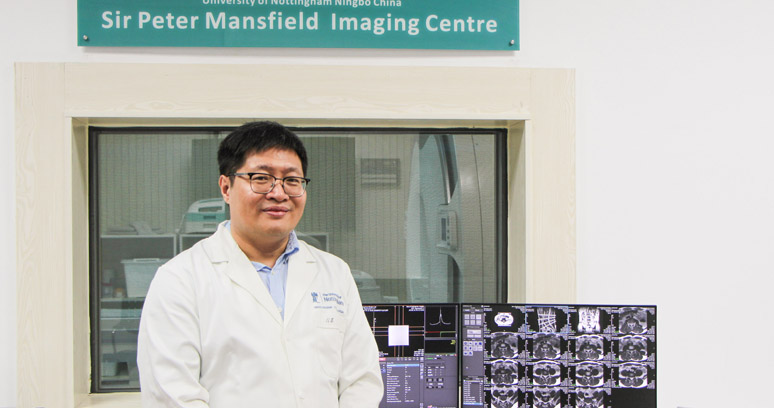
Associate Professor in Magnetic Resonance Imaging
Can you explain your research?
My expertise lies in biomedical engineering, especially the magnetic resonance imaging (MRI). My work focuses on the development of biomedical imaging techniques and hardware to meet people’s needs in improving health and wellbeing.
I returned to China in 2013 to join the University of Nottingham Ningbo China (UNNC). With the joint effort of the University, we brought the world-renowned Sir Peter Mansfield Imaging Centre from the UK campus to UNNC and established its Ningbo branch centre, where my team does the majority of our research work.
To explain our research, it might be easier to draw an analogy between the MRI machine and a camera. A camera uses light to form pictures, and MRI employs magnets to produce magnetic images of the organs in the body. What we do is to create more economical and flexible MRI machines that could generate clearer images, and to develop more functionalities. Our work could help hospitals solve practical problems in examining and treating diseases.
What challenges is your research hoping to tackle? How will your research affect the average person?
Our research focuses on solving clinical problems from both the technical and hardware aspects of MRI.
One of our projects, in collaboration with Moscow State University and an Indian hospital, is to employ multinuclear MRI technologies and hyperpolarised noble gas for lung MRI diagnosis.
In China, we have the world's largest population of smokers, and lung disease has long been a problem to Chinese people. Over the last century, there has been no substantial progresses in the treatment of lung diseases, and the mortality rate remains high.
Although CT is commonly used in hospitals as the "golden standard" for diagnosing lung disease, it produces harmful ionizing radiation. For patients recovering from COVID-19, who need health assessments every three months, the use of CT is not only damaging to the body, but also ineffective in providing diagnostic information.
What we aim to do in this project is to enhance MRI imaging for the use of patients’ respiratory function and blood perfusion investigation.
Sir Peter Mansfield Imaging Centre at UNNC
The other project we do is to upgrade the hardware. Existing MRI device relies heavily on liquid helium as the refrigerant for superconducting magnets, which has several major drawbacks. Helium is a natural scarcity, and MRI devices using liquid helium can only work when patients lying still. Another drawback is that it requires continuous refrigeration to keep helium in the liquid state, which can be a major safety hazard.
We are now collaborating with a manufacturer in Yuyao and Ningbo’s hospitals to conduct research on the development of cryogen-free superconducting magnets, using cheaper and safer copper tape as an alternative to liquid helium.
What are the achievements so far?
For the hyperpolarised noble gas project, our team has successfully applied hyperpolarised noble gas such as helium3, xenon129 and krypton83 to the imaging process and developed an advanced MRI technique that could form pictures of the airspace of lungs.
This technology will enable the early detection of lung diseases in an accurate but non-invasive way.
The cryogen-free MRI project is now supported by the Ministry of Science and Technology. A whole-body cryogen-free MRI that we developed has been running steadily for one year, proving the feasibility of this technology.
This breakthrough has also made it possible to create rotatable MRI devices, which would allow patients to be scanned and diagnosed in a standing position. We are currently developing a 1.5T rotatable dual gesture cryogen-free MRI, which should be the world's first clinically available whole-body rotatable MRI. We expect this machine to be set up at UNNC in the coming year.
In addition, our team is also developing mobile MRI, silent MRI, MRI for infants, and more.
We understand that your team's research covers a wide spectrum of topics. In addition to clinical applications, you are exploring applications in the biological area. Can you share more on that aspect?
We are exploring the use of MRI techniques for rapid food quality assessment. Taking fruit for instance, MRI can evaluate a series of quality indicators, such as the freshness, sugar content, and pesticide residue. In terms of the speed of detection, if we take orange as an example, our test can scan more than 1,000 oranges in a minute for sugar content. We expect that once the technology gets commercialised, the cost could be down to about 5 cents per orange. The use of MRI for food quality control is promising.
Can you give an introduction of your research team?
We have Professor Thomas Meersmann, who is the Li Dak Sum Chair Professor in Translational Imaging on the team. Professor Meersmann and I are just about to do an in-depth research together on the hyperpolarised noble gas project. We also have Dr Jing Wang on the team, who is an expert on optical sensors, and Dr Shun Bai, who specialises in electromagnetism and radio frequency radiation.
In addition, we have six PhD students. Several of them are currently developing the MRI technique for the use of detecting blood perfusion in lungs, livers and other body parts. The project should advance to the clinical study phase later this year with Ningbo No.2 Hospital.
Watch a video